Helen, 49 years old female patient came to Kokilaben D Ambani Hospital with upper backache since 8 years on and off. No history of weakness or paresthesia. Neurologically there was no deformity or tenderness. MRI spine showed a large solid heterogeneously enhancing tumour of size 8.5 x 8.0 x 10 cm arising from right sided Thoracic (T6) nerve root with widened neural foramina causing displacement of spinal cord to left with mild compression.
MRI

Fig 1 – MRI sagittal view of complete spine showing Intradural component of tumour at T5-T6 level
Fig 2- MRI Spine coronal view showing hyperintense tumor in posterior mediastinum extending from T2 to T9 level on right side.

Fig 3 - MRI Spine coronal view showing heterogenous enhancement of tumour with dumbbell shaped tumour causing widening of neural foramina.

Fig 4 - MRI Spine axial view showing heterogenous enhancement of tumour with dumbbell shaped tumour causing widening of neural foramina.

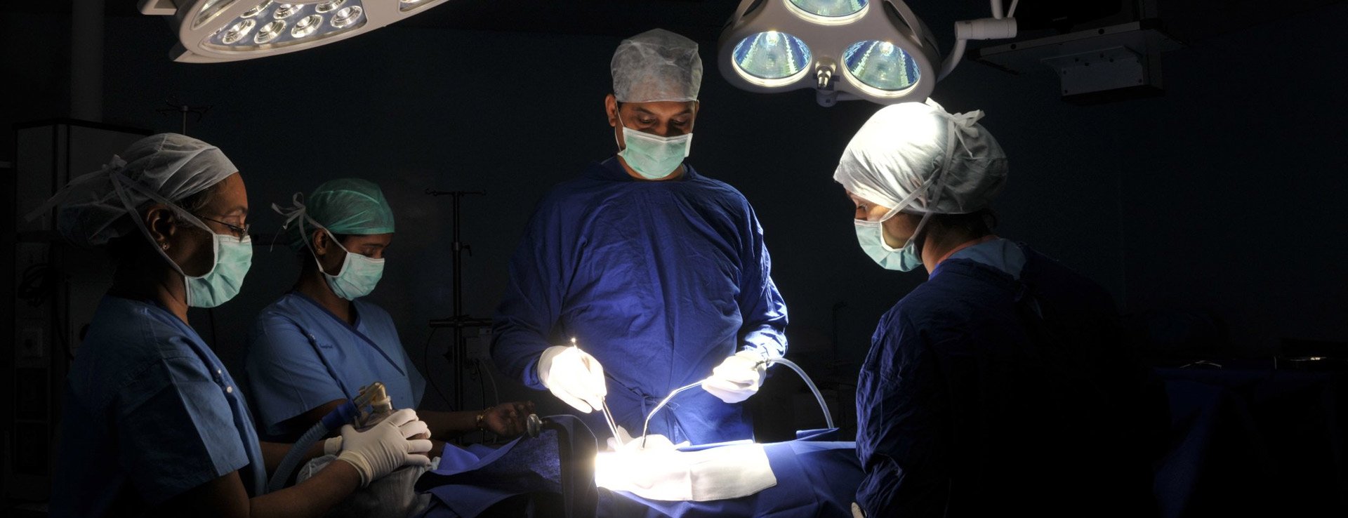
She underwent Right sided thoracotomy with excision of mediastinal mass and intradural T5-T6 neurogenic tumour by Dr Abhaya Kumar on 28/12/2016.
Postoperatively, ICD was removed and patient was discharged on 10th day without any deficit and total tumour excison.
Histopathology report showed bening schwannoma.
Stay updated to all the latest news and offers at KDAH
