There is a severe shortfall of genuine vascular care in our country. At Kokilaben Hospital, we offer authentic comprehensive vascular care through a world-class facility.
Aneurysm is ballooning out or bulging of the wall of the blood vessel. This occurs because of weakening of the wall of the blood vessel. It may involve the whole circumference or only a localised segmental wall of the blood vessel.
Wall of the blood vessel consists of three layers:
Inner: Intimal lining
Middle: Thick muscular layer
Outer: Fibrotic attachment – Adventitia. It is the middle layer which becomes weak
The flowing blood inside the blood vessels exerts pressure on the weak wall and leads to aneurysm formation.
Aorta is the main blood vessel distributing blood to all parts of the body. It carries blood at a high pressure: i.e. 120/80 mm of mercury, in a normal individual.
When the aorta starts dilating or bulging, it is called Aortic Aneurysm. When the aorta in the chest enlarges, we call it Thoracic Aortic Aneurysm (TAA) and in the abdomen, it is called Abdominal Aortic Aneurysm (AAA).
There are known risk factors which lead to development of Aortic Aneurysm.
These are:
In majority of cases, aneurysm is detected incidentally, during test like x-ray of the chest, abdomen, or spine. Ultrasonography or CT/MRI can detect aneurysm.
However, few patients can have symptoms like Thoracic Aortic Aneurysm:
Abdominal Aortic Aneurysm
Loss of consciousness drops in blood pressure, sweating, and distension of abdomen signifies rupture of aneurysm.
Rupture of the aneurysm leads to sudden leak of blood, which leads to drop in blood pressure and loss of blood. This is associated with high risk of death and poor health. This event occurs rapidly and often, patients cannot survive and may not reach the hospital.
People who are at high risk of developing Aortic Aneurysm should undergo regular check-up like x-ray chest and/or ultrasonography. In fact, every male over 65 years of age should undergo sonography. One should demand for screen for Aortic Aneurysm when you go for sonography/x-ray chest or general body health check up program.
Diameter size of the aortic aneurysm is the main criteria to decide treatment of the size of the aorta becomes double the size of normal aorta, then this aneurysm needs treatment or else it is at higher risk to rupture.
In the thorax: If the aneurysm is > 5.5 cm in size.
In the abdomen: If the aneurysm is > 5 cm in size.
In some patients and conditions even a smaller size of the aneurysm needs urgent treatment.
Presence of calcium, some symptoms, eccentric bulge, and infective aneurysm with aneurysm in females may need early treatment.
When Aortic Aneurysm is detected it is important to meet a doctor. Control of blood pressure, avoiding or countering the possible risk factors and avoiding exertion and jerks in the chest, abdomen with back are some of the standard care. If the aneurysm is smaller than the critical size then a close check up every 6 months is necessary. USG/CT scan can be undertaken to monitor the size.
If these care and precautions are ignored then there is high risk of rupture of the aneurysm. This is associated high morbidity, mortality, and cost.
Critical size:
Thoracic Aorta = 5.5 to 6 cm
Abdomen Aorta = 5 cm
Aneurysm larger than this size needs urgent interventions such as:
Surgical repair
Endovascular stent graft
Hybrid-surgery + Endovascular
These are complex procedures, needs general anaesthesia, stay in the ICU and hospital. Complications and death risk are 5 to 7 per cent in most of the cases.
Lumen of the blood vessel reduces with deposition of cholesterol (fatty substance) on the inner lining of the blood vessel. This reduces the flow of blood in the vessel and supply of oxygen to the tissue.
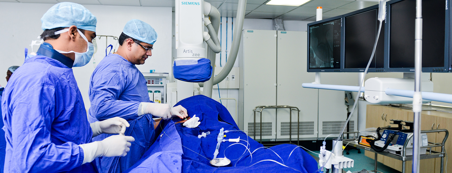
Angioplasty is a procedure wherein a catheter with a balloon is introduced at the site of blockage. With inflation of the balloon, the plaque is compressed and fragmented. This improves the luminal diameter of the blood vessel and hence the blood flow.
Stent is a mesh like metallic spring like tube. Stent scaffolds the blood vessel wall and prevents the blood vessel from re-blocking.
Stent-grafts are specialised medical devices which a metallic spring like device, this is a medical fabric stitched. This forms a closed tube-a new tube within the aorta. Outside the graft, blood does not flow and forms a clot. Inside the stent graft, blood flows normally. Hence, the stent graft actually excludes the aneurysm. Hence, the risk of rupture of the aneurysm reduces. Over a period the dilatation of the aorta remodels and reduces in diameter.
Based on the CT angiography findings, we as Interventional Radiologist plan and decide the treatment.
The advantages of endo-vascular treatment are:
Cost varies depending on the anatomical findings on CT scan. The average cost of the stent graft is Rs. 4.5 lakhs to 6.0 lakhs. Added to this is the cost of hospitalization, OT/ICU and catheter lab. Other consumables had doctors fee would vary.
To detect Aortic Aneurysm ultrasonography, CT scan or MRI studies are performed at Radiology Clinics. However, qualified and trained Interventional Radiologist is capable of guiding patients in the management of Aortic Aneurysm. Other specialist like vascular surgeons, CVTS surgeon and some cardiologists too treat Aortic Aneurysm.
Diabetic Foot, DVT Management, Aortic Disease, Liver Tumour Management, Interventions in Women Health.


