Obstetric Ultrasound and Fetal Medicine is a rapidly emerging specialization in Obstetrics that enhances Maternity care and optimizes outcomes in both - High Risk pregnancies as well as apparently low risk ones. At Kokilaben Hospital, we have a qualified specialist in this area and the services we aim to provide are detailed below.
The First Trimester Screening or Nuchal Translucency Scan or Nuchal Scan
This scan is carried out from 11 weeks to 13 weeks and six days of pregnancy.
This scan aims to look at viability, dating, assess the risk of having a baby with a chromosomal abnormality leading to mental handicap (Down Syndrome), assess the risk of having a baby with a cardiac defect and checks the babys anatomy (early check).
The vast majority of babies are normal. However all women, whatever their age, have a small risk (1-2%) of delivering a baby with a physical and/or mental handicap. Down Syndrome is the commonest chromosomal abnormality in liveborn babies (1:900). Although the risk of having a Down Syndrome baby increases with advancing maternal age, all pregnancies are at some risk and it is vital in this modern age that the basic screening test is offered to all pregnant women.
The most accurate way of estimating the risk of the Fetus having Downs Syndrome is carried out at 11-13 weeks and depends on the:
After the scan and the blood test, on the basis of all the above factors, the estimated risk for Downs Syndrome will be discussed. Further decisions regarding invasive diagnostic tests (amniocentesis or CVS) will be discussed with the referring Obstetricians, if they wish to do so. Irrespective of whether or not an invasive test is performed, it is recommended that a scan is performed at 19-20 weeks to check for physical abnormalities (detailed anatomy or anomaly scan).
It is important to have this scan and risk assessment done by sonographers / obstetricians/radiologists who are correctly trained and certified in this technique and are using the software that is of a high standard. The Fetal Medicine Foundation, London is the accepted authority in the world for the training, certification and licensing of the software for risk calculation.
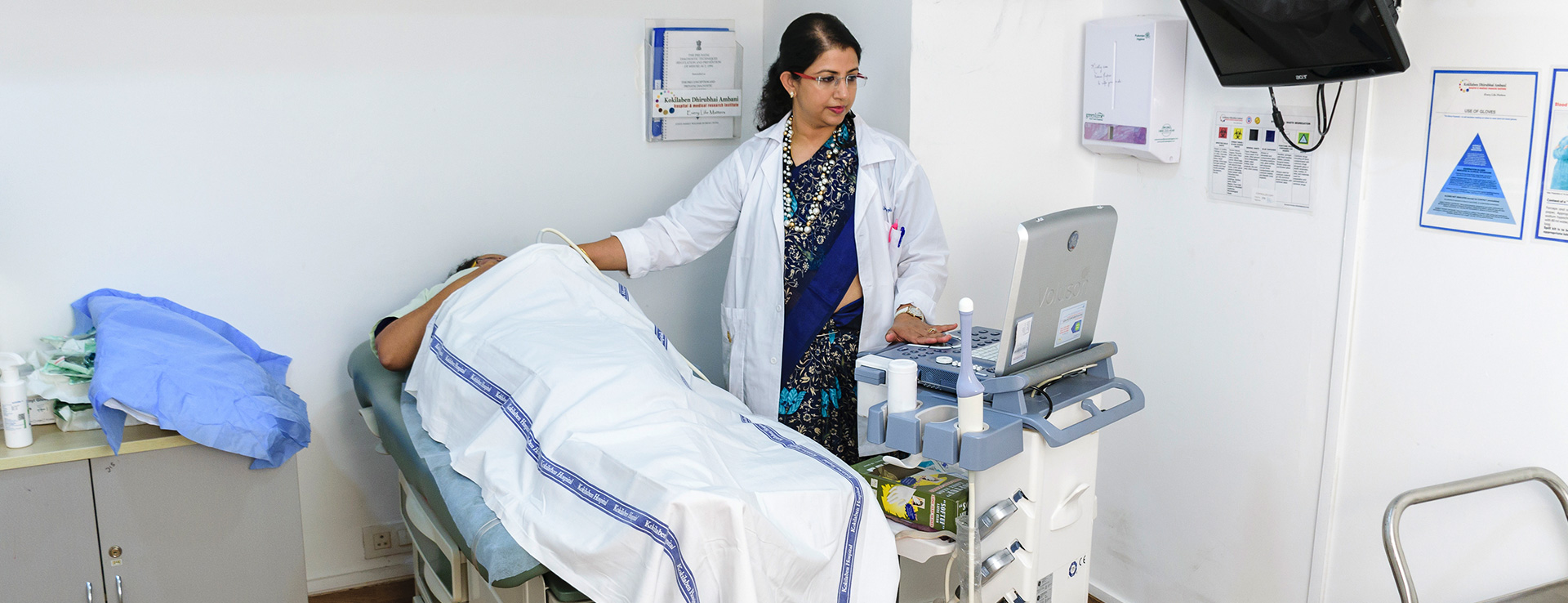
At Kokilaben Hospital, we have a Fetal Medicine Foundation certified specialist, who performs this scan and further tests, if necessary - Dr. Shantala Vadeyar
This is a detailed scan at 18-20 weeks of pregnancy.
During the scan we examine each part of the fetal body, determine the position of the placenta, assess the amount of amniotic fluid, and measure fetal growth. Special attention is paid to the brain, face, spine, heart, stomach, bowel, kidneys and limbs. In women at high risk for preterm delivery (multiple pregnancies, previous preterm birth, abnormalities of the uterus or previous cervical surgery) we also carry out a transvaginal scan to measure the length of the cervix.
If any abnormalities are detected, the significance of the findings will be discussed with the referring Obstetrician and the couple will be given the opportunity to have further counselling.
A detailed examination of the fetal heart and connecting vessels is carried out usually at 20 - 22 weeks.
This ultrasound scan is usually carried between 28 - 39 weeks of pregnancy. Some Obstetricians advise that this scan is offered to all women. Others reserve such scans for those women who have had previous complications of pregnancy such as pre-eclampsia, growth restriction, diabetes, stillbirth, and for those women who develop a problem during the course of their current pregnancy.
This scan aims to determine the growth and health of the fetus by:
These scans are getting very popular among couples as a way of bonding with their baby. They are best performed between 28-34 weeks, while the babys head is not fixed in the pelvis. An adequate amount of amniotic fluid / liquor is required to be present around the babys face and the position of the baby needs to be suitable for good visualisation for 3D/4D scans. Sometimes, this cannot be guaranteed and one has to accept these limitations!


