Best Oesophagus Cancer Treatment in Mumbai

Cancer Oesophagus

The Oesophagaus is a tube that starts in the neck, traverses through the chest and joins the stomach in the upper abdomen. It transports food from mouth into the stomach. In cancer of the oesophagus (food pipe), malignant (cancerous) tumour arises in the innermost lining of the tube. It then progresses outwards, lengthwise and circumferentially to cause progressively increasing difficulty in swallowing. Two most common form of cancer oesophagus are squamous cell carcinoma and adenocarcinoma; latter generally involved lower part of the oesophagus near stomach.

Tobacco and heavy alcohol use are known to increase the risk of developing cancer of the oesophagus. People with long standing reflux disease and anaemia (women in particular) also have increased risk of developing cancer of the oesophagus.

Warning signs

  • Difficulty in swallowing/ painful swallowing
  • Weight loss
  • Pain behind the breastbone
  • Hoarseness and cough
  • Indigestion and heartburn
  • Black stools or blood in vomit
  • Long standing reflux (acidity)
  • Tobacco, alcohol and tambul (betel nut)
  • Gastroesophageal reflux disease (GERD)
  • Obesity
  • Exposure to chemical fumes
  • Injury to oesophagus

Progression

  • Stage 0: Cancer is confined to the lining of the oesophagus
  • Stage 1: Cancer has spread deeper into the tissues of the oesophagus
  • Stage 2: Cancer may now affect lymph nodes near the oesophagus
  • Stage 3: Cancer has progressed to surrounding tissues
  • Stage 4: Cancer has metastasised, or spread, to other parts of the body

Diagnosis

  • Primary diagnosis: Physical examination
  • Secondary diagnosis: Barium swallow, Oesophago-Gastro-Duodenoscopy, CT Scan with contrast, PET CT scan whole body

The following tests and procedures are necessary for confirming the diagnosis:

  • Barium Swallow: A series of x – rays of the esophagus and stomach. The patients drinks a liquid that contains barium (a silver white metallic compound). The liquid coats the esophagus and x ray is taken.
  • Oesophago-Gastro-Duodenoscopy: This involves introducing a flexible telescope through the mouth to visualize esophagus and stomach. A biopsy may be done at the same time if tumour is seen at endoscopy.
  • CT Scan with Contrast: This is computerized imaging of chest and abdomen done after intravenous and oral contrast. This gives information about the location, extent and spread of spread disease and hence helps in planning treatment.
  • PET CT Scan Whole Body: This is combination of CT scan entire body and PET. It involves giving a special dye before scanning. The dye (FDG) helps in identifying the spread of disease and thus is more accurate for staging of disease and planning of treatment. The treatment depends on the stage of cancer. Cancer of the esophagus is staged from I to IV; stage I is early, localised cancer and stage IV is when cancer has spread to other organ such as lung, liver bone, etc., or neighbouring structures, such as tracheo-bronchial tree, nerves or aorta.
The most common signs of esophageal cancer are painful or difficult swallowing and weight loss. A doctor should be consulted if any of the following problems occur:
  • Painful or difficult swallowing
  • Weight loss
  • Pain behind the breastbone
  • Hoarseness and cough
  • Indigestion and heart burn
  • Black stools or blood in vomit

The following tests and procedures are necessary for confirming the diagnosis:

  • Barium Swallow: A series of x – rays of the esophagus and stomach. The patients drinks a liquid that contains barium (a silver white metallic compound). The liquid coats the esophagus and x ray is taken.
  • Oesophago-Gastro-Duodenoscopy: This involves introducing a flexible telescope through the mouth to visualize esophagus and stomach. A biopsy may be done at the same time if tumour is seen at endoscopy.
  • CT Scan with Contrast: This is computerized imaging of chest and abdomen done after intravenous and oral contrast. This gives information about the location, extent and spread of spread disease and hence helps in planning treatment.
  • PET CT Scan Whole Body: This is combination of CT scan entire body and PET. It involves giving a special dye before scanning. The dye (FDG) helps in identifying the spread of disease and thus is more accurate for staging of disease and planning of treatment.
The treatment depends on the stage of cancer. Cancer of the esophagus is staged from I to IV; stage I is early, localised cancer and stage IV is when cancer has spread to other organ such as lung, liver bone, etc., or neighbouring structures, such as tracheo-bronchial tree, nerves or aorta.

Stage I/II

  • Surgery is the treatment of choice in medically fit person.
  • Radiation+Chemotherapy are usually prescribed in patients who cannot with stand major surgery due to coexisting medical illnesses or for those who are not willing for surgery.

Stage III

Induction Treatment Followed By Surgery: Downstaging of disease is done with chemotherapy alone or combination of chemotherapy and radiation therapy. Later may be associated with increased side effects. This is followed by reassessment of the disease status. If adequate downstaging is achieved surgery is preferred.

Radiation Combined With Chemotherapy: Those patients who are medically unfit to go through surgery are treated with combination of radiation therapy and chemotherapy or radiation alone.

Stage IV (Presence of Metastasis)

  • Stenting with self expanding stent
  • Palliative radiation therapy
  • Palliative chemotherapy
  • Supportive care alone – for very sick patients whose life expectancy is few weeks

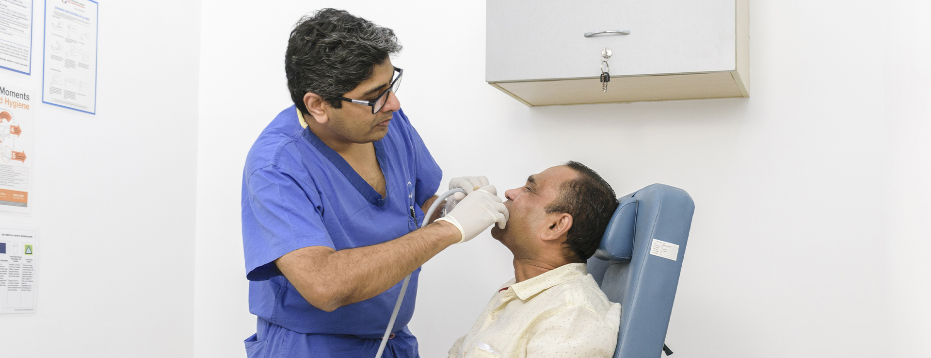
In early stages when the cancer is very superficial endoscopic resection of disease (EMR - Endoscopic Mucosal Resection) can be done.

Surgery

Surgery entails removal of oesophagus (subtotal or total) along with adjacent lymph nodes. This can be the done using the conventional open technique or by using minimal invasive technique (key hole surgery). Later procedure can be performed using the assistance of da Vinci Robot. Minimal invasive surgery is associated with faster recovery and small incisions.

After surgically removing the oesophagus, stomach is made into a tube and brought up in the neck or chest and anastomosed (joined) to the proximal cut end of the oesophagus. The usual stay in hospital is for about 10 to 12 days.

Radiation Therapy

In radiation therapy ionising rays are targeted on to the tumour and adjacent area of interest using linear accelerator. It damages cells within the field of radiation; cancer cell cannot recover after damage but normal cells recover and function as before. Radiation is given in small fraction daily, five days a week (Monday to Friday) over a period of 4-6 weeks. In patient who are fit chemotherapy may be given along with radiation (once every week or at three weekly interval) to enhance the efficacy of radiation therapy.

Chemotherapy

Chemotherapy consists of giving anti cancer medication to target cancer cells. Generally combination of two or three drugs is given along with saline every three weeks. Each course may be instituted over three days. Anti cancer medication target all rapidly multiplying cells in the body; cancer cells multiply rapidly and hence are targeted and destroyed; however only a fraction of cancer cells get killed with each course. Normal cells which multiply rapidly also get affected by these drugs. However, these recover unlike cancer cells, and thus, side effects are generally temporary. Medication is also given during the chemotherapy to reduce associated side effects. Chemotherapy helps in reducing the cancer burden and down staging the disease when given before definitive surgery; occasionally (10%) it may succeed completely eliminating the cancer.

Stenting

In patients with advanced disease where cure is not possible (stage IV) or those who are elderly and unfit for definitive treatment swallowing can be restored by deploying Self Expanding Stent (SEMS). This is done endoscopically by a gastroenterologist. Patient will be able to swallow liquids and semisolids after stenting which remains in oesophagus till the end.Stenting restores swallowing alone and is not an alternative to curative treatment.
Amongst many other factors, the stage of cancer at the time of diagnosis is the most important determinant for cure. Cure rates are best when tumour is localised and superficial without having spread to lymph glands. The risk of recurrence of cancer increases with advancing stage. In stage IV, where cancer has already spread to other organs, disease is not curable. The risk of recurrence after successful treatment is maximum during the first two years; after 5 years the risk becomes negligible.

Difficulty in swallowing is the most common complain with which patient present. Difficulty in swallowing occurs when lumen of the oesophagus is almost completely blocked; this occurs very late in the course of disease. Early symptoms such as long standing retrosternal burning, painful swallowing and blood in vomiting should not be ignored and investigated by endoscopy.

Milestones

90 per cent of oesophageal cancer surgeries at Kokilaben Hospital are done using Video-Assisted Thorocoscopic Surgery (VATS). VATS is a keyhole surgery that reduces postoperative pain, aids in early recovery, avoids prominent scars, and reduces length of hospital stay.

Top tips

  • Quit smoking
  • Stop drinking alcohol, or try to cut back
  • Consult a doctor if you experience persistent heartburn that may be a sign of gastroesophageal reflux disease (GERD)
  • Commit to a regular exercise program and avoid becoming overweight.

Remarkable Case

Successful Surgery to Remove the Largest Mediastinal Tumour, Weighing 7.5 Kg

Background and diagnosis : A 19-year-old student was suffering from difficulty in breathing at rest and inability to sleep. He was investigated outside and diagnosed with a huge tumour in the chest (mediastinal teratoma). He was treated with three courses of chemotherapy with no response. On examination, he was breathless and unable to lie down even for the examination. The apex beat was palpable in the right chest in the 5th intercostals space. A CT scan revealed a mass occupying the entire left chest with shift of the heart to the right.

Treatment : After investigations and high-risk consent he underwent surgery. The tumour was exposed through a T-incision consisting of median sternotomy and left-sided antero-lateral thoracotomy. It was fully removed along with the entire left lung. The patient was electively ventilated for 48 hours.

Outcome : Postoperative recovery was largely uneventful and the patient was discharged on the 15th day after the surgery.

Dr. Rajesh Mistry -  Best oncologist Near Me

Dr. Rajesh Mistry

MBBS, MS

Department

Cancer/Surgical Oncology, Robotic Surgery, Minimal Access Surgery

Expertise

Esophageal surgery: VATS and Robotic; Pulmonary surgery (Malignant and Benign): VATS and Robotic; Cancer of Thymus and other mediastinal masses; Chest wall tumours; Gastric Cancer; Head Neck Oncology